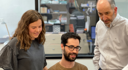
Researchers of LPI create 'synthetic' images that could improve the diagnosis of malignant brain tumours

Artificial Intelligence has enabled two pre-doctoral researchers at the University of Valladolid, Rafael Navarro and Elisa Moya, to generate synthetic images that can be used in combination with those adquired in an MRI scaner in a radiomic system for survival prediction of patients with glioblastoma. Glioblastoma is an aggressive and fast-growing brain tumour, comprising the most common type of malignant brain tumour, with a survival rate of approximately 40% in the first year after diagnosis and 17% in the second year, so predicting its survival is key for efficient treatment and surgery planning.
This new technique adds to those currently used in the diagnosis, prognosis and therapeutic response to this brain cancer, using synthetic magnetic resonance imaging and radiomics. "The synthetic images are generated with an artificial intelligence system that has been trained from a large number of real images obtained on MRI scanners. We then use mathematical measures to compare the synthetic images with the real ones and, in addition, these images are used in the radiomic system, which is responsible for extracting quantitative characteristics from the images in order to achieve a predictive tool for this type of brain cancer. This can allow better planning of treatment or surgery," explain the young scientists.
The generation of these images will also make it possible to reduce the duration of MRI scans, replace artifacted images, and generate databases that help in the diagnosis of the disease, as Elisa Moya points out, "During the adquisitions of the MR images, the patient must remain completely still, which for some people, with claustrophobia problems, or children, for example, is quite uncomfortable. This new technique gives them higher comfort and also reduces costs, since we would only need to adquire two magnetic resonance images and, then, the other ones can be synthesized, so the time in the scanner would be reduced.
The study is carried out by Rafael Navarro, Biomedical Engineer at the Polytechnic University of Madrid, who is doing his PhD "Radiomics in neuroimaging: use of magnetic resonance features for the prediction of results in brain pathology", at the LPI; in collaboration with Elisa Moya, a telecommunications engineer from the University of Valladolid who is also developing her PhD thesis ("Obtaining parametric maps of T1, T2 and DP from routine MRI sequences using artificial intelligence") in this laboratory.
The work, published in the scientific journal "NMR in Biomedicine", has been led by Dr. Rodrigo de Luis, Dr. Santiago Aja and Dr. Carlos Alberola. Funding to support this work comes from the Asociación Española Contra el Cáncer and the Ministerio de Ciencia e Innovación of Spain.
- Link to the journal paper: https://analyticalsciencejournals.onlinelibrary.wiley.com/doi/full/10.10...
- Link to 1-minute power speech video (in Spanish): https://www.youtube.com/watch?v=nr-O8ehgs90&t=2s
- Link to an article in the local media (in Spanish): https://www.elnortedecastilla.es/valladolid/imagenes-sinteticas-luchar-2...
